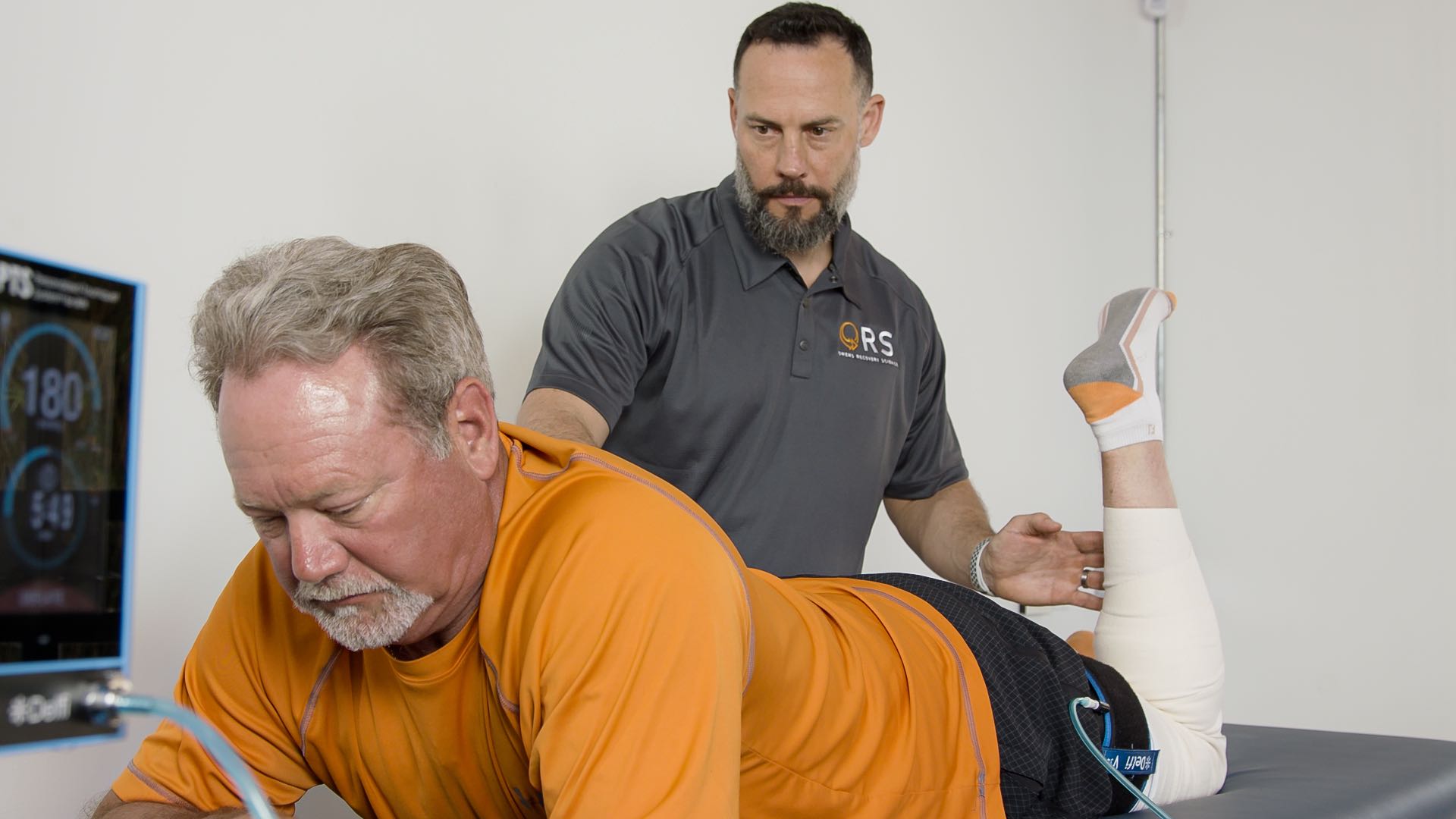
A potential risk for clotting or venous thromboembolism (VTE) has been a commonly voiced concern since the early days of BFR application. This is an understandable concern given the publications that highlight an increased incidence rate of VTE after surgery, and many common orthopedic procedures involve tourniquet use. (Sweetland et al. 2009)
This raises a few questions:
- What causes VTE?
- Does the application of a tourniquet for surgery increase the incidence of VTE post-surgically?
- Would the use of a tourniquet for BFR increase risk of VTE?
The formation of a VTE appears to be multifactorial. Bond et al recently put out a very good review in which they discuss a balance between coagulation and fibrinolysis. (Bond et al. 2019) They highlight that the formation of a VTE typically comes from a combination of more than one of the following: inherited or acquired thrombophilia (hypercoagulability), endothelial injury, and stasis. (Bond et al. 2019) These three factors are referred to as Virchow’s triad and should be screened by the clinician. Endothelial injury is implied following orthopedic surgery, and will be coupled with physician imposed or self-imposed activity limitation (stasis). Clinically we should be mindful of the signs and symptoms of DVT: unexplained swelling, pain, cramping, soreness, red or discolored skin, or feeling of warmth in the affected limb. Additionally, some useful tools like this one from Caprini have been developed: https://venousdisease.com/dvt-risk-assessment-online/
According to a study by Ocak et al, the incidence for VTE is around 1 to 2 per 1000 people per year. (Ocak et al. 2013) That’s only a 0.1 to 0.2% incidence rate which is pretty low, albeit higher than many clinicians might have expected. This risk is significantly amplified after surgery. A large scale study by Sweetland et al followed 947,000+ middle aged women in the UK and found they were 70 times more likely to have a VTE in the 6 weeks following an inpatient surgery and 10 times more likely in the 6 weeks after an outpatient procedure.(Sweetland et al. 2009) Further, studies have sought to determine the causality of VTE following surgery with or without tourniquet use. A study by Jarrett et al found a high incidence of asymptomatic VTE intra-operatively that was not statistically different between tourniquet and non-tourniquet groups. (Jarrett et al. 2004) Fascinatingly, they reported visualized asymptomatic clots via ultrasound in 70% of the individuals. There is no question the risk for VTE is elevated during and after surgery, but it doesn’t appear that the application of a tourniquet is directly related to the elevated risk. Interestingly, Jarrett et al went on to say, “It is possible that small venous emboli are a common physiological event during life and that the lungs are readily able to clear small numbers of small emboli.” (Jarrett et al. 2004)
The risk for VTE with BFR has been assessed both acutely and chronically; we can also look at reported events longitudinally. Some of the common blood markers for VTE include D-Dimer, Fibrinogen, and CRP. BFR studies on young healthy individuals, individuals after bed rest or at simulated altitude, elderly individuals, or individuals with ischemic heart disease found no elevation in markers for VTE after one application. (Manini et al. 2012; Madarame et al. 2010; Nakajima et al. 2007; Fry et al. 2010; Madarame et al. 2013) Studies that assessed markers for VTE across multiple weeks of BFR application in young healthy individuals, elderly individuals, and after knee arthroscopy also found no elevation compared to a work-matched control. (Clark et al. 2011; Yasuda et al. 2014; Yasuda et al. 2014; Tennent et al. 2017) Johnny Owens and his co-authors at the Department of Defense examined bilateral duplex ultrasound evaluations on subjects using BFR or work matched free-flow controls before surgery and 4-weeks after knee arthroscopy and found no differences between groups. When looking at the totality of the research, there is one reported case of an individual performing chronic BFR and a VTE episode. (Noto et al. 2017) The case report is on a female instructor for KAATSU that had been performing bilateral UE BFR 3 days a week for 2 years with 30 minutes to an hour inflation of the cuffs per session. She reports having localized swelling and tenderness around the left collarbone for the 6 years leading up to the VTE. She was diagnosed with Thoracic Outlet and Paget-Schroetter Syndrome. Even though the application of BFR, in this case, doesn’t match current guidelines, it’s hard to say that BFR is what caused the VTE in light of her chronic confounding symptoms.
Further reassurance can be drawn from the fact that across millions of clinical applications there are not any reported VTE events involving BFR. Our recommendation will always be to apply BFR in a way that matches up with current best practices. If a patient doesn’t exhibit any signs or symptoms of a VTE, there is a very low likelihood that BFR will increase the risk. As with everything done in a rehabilitation setting, sound reasoning and good clinical judgement are important for safe and successful implementation of BFR.
Bond et al described key points for best VTE practice that align 100% with the guidelines we at Owens Recovery Science have championed:
“Some clinicians may use rudimentary techniques to achieve occlusion, such as surgical tubing or elastic straps wrapped tightly around the proximal portion of the exercising limb. This method may or may not completely occlude all arterial and venous blood flow, and there is no way of knowing what occlusive pressure the vessels experience. Further, the relatively thin diameter of the tubing or straps may cause highly localized stresses and an ineffective transmission of pressure, potentially causing direct damage to the soft tissues and structures underneath the application site”.
“A pressure specific to each individual would be the safest prescription method, because the same pressure may not necessarily occlude the same amount of blood flow for all individuals under the same conditions”. ie...avoid unnecessary stasis
“The cuffs developed for BFR are wider than those traditionally used. The most frequently used cuff width is 10 to 12 cm, although cuffs greater than 15 cm may be more desirable. Additionally, cuffs are now tapered to conform with the natural proximal-to-distal narrowing of the thigh or upper arm and are limb-circumference specific, which allows them to be fitted to various limb circumferences. Together, these advances enhance the transmission of pressure, and may appropriately mitigate complications caused by finely localized stresses from the cuff itself and reduce the pressure required to reach the same level of occlusion achieved with narrower cuffs”.
“The collective literature suggests that a proper prescription of BFR in the context of Virchow’s triad would not heighten the risk of developing VTE. Nonetheless, clinicians need to thoroughly screen for VTE signs and symptoms and be cognizant of each patient’s VTE risk factors before proceeding with BFR, particularly given the risk of asymptomatic DVT. Health care professionals must also make sure they have the proper training and are using the correct BFR equipment and pressure prescription techniques to ensure their patients’ safety”.
- Sweetland S, Green J, Liu B, et al. Duration and magnitude of the postoperative risk of venous thromboembolism in middle aged women: prospective cohort study. BMJ. 2009;339:b4583.
- Naess IA, Christiansen SC, Romundstad P, Cannegieter SC, Rosendaal FR, Hammerstrøm J. Incidence and mortality of venous thrombosis: a population-based study.
- J Thromb Haemost. 2007;5(4):692-699. Ocak G, Vossen CY, Verduijn M, et al. Risk of venous thrombosis in patients with major illnesses: results from the MEGA study: Major illnesses and venous thrombosis. J Thromb Haemost. 2013;11(1):116-123.
- Bond CW, Hackney KJ, Brown SL, Noonan BC. Blood Flow Restriction Resistance Exercise as a Rehabilitation Modality Following Orthopaedic Surgery: A Review of Venous Thromboembolism Risk. J Orthop Sports Phys Ther. 2019;49(1):17-27.
- Nakajima T, Takano H, Kurano M, Iida H. Effects of KAATSU training on haemostasis in healthy subjects. of KAATSU Training …. 2007. https://www.jstage.jst.go.jp/article/ijktr/3/1/3_1_11/_article/-char/ja/.
- Jarrett PM, Ritchie IK, Albadran L, Glen SK, Bridges AB, Ely M. Do thigh tourniquets contribute to the formation of intra-operative venous emboli? Acta Orthop Belg. 2004;70(3):253-259.
- Madarame H, Kurano M, Fukumura K. Haemostatic and inflammatory responses to blood flow‐restricted exercise in patients with ischaemic heart disease: a pilot study. Clin Physiol. 2013. http://onlinelibrary.wiley.com/doi/10.1111/j.1475-097X.2012.01158.x/full.
- Yasuda T, Fukumura K, Uchida Y, et al. Effects of Low-Load, Elastic Band Resistance Training Combined With Blood Flow Restriction on Muscle Size and Arterial Stiffness in Older Adults. J Gerontol A Biol Sci Med Sci. 2014. doi:10.1093/gerona/glu084
- Yasuda T, Fukumura K, Fukuda T. Muscle size and arterial stiffness after blood flow‐restricted low‐intensity resistance training in older adults. J Med. 2014. http://onlinelibrary.wiley.com/doi/10.1111/sms.12087/full.
- Fry CS, Glynn EL, Drummond MJ, et al. Blood flow restriction exercise stimulates mTORC1 signaling and muscle protein synthesis in older men. J Appl Physiol. 2010;108(5):1199-1209.
- Clark BC, Manini TM, Hoffman RL, et al. Relative safety of 4 weeks of blood flow-restricted resistance exercise in young, healthy adults. Scand J Med Sci Sports. 2011;21(5):653-662.
- Madarame H, Kurano M, Takano H, et al. Effects of low-intensity resistance exercise with blood flow restriction on coagulation system in healthy subjects. Clin Physiol Funct Imaging. 2010;30(3):210-213.
- Manini TM, Yarrow JF, Buford TW, Clark BC, Conover CF, Borst SE. Growth hormone responses to acute resistance exercise with vascular restriction in young and old men. Growth Horm IGF Res. 2012;22(5):167-172.
- Tennent DJ, Hylden CM, Johnson AE, Burns TC, Wilken JM, Owens JG. Blood Flow Restriction Training After Knee Arthroscopy: A Randomized Controlled Pilot Study. Clin J Sport Med. 2017;27(3):245-252.
- Noto T, Hashimoto G, Takagi T, et al. Paget-Schroetter Syndrome Resulting from Thoracic Outlet Syndrome and KAATSU Training. Intern Med. 2017;56(19):2595-2601.


