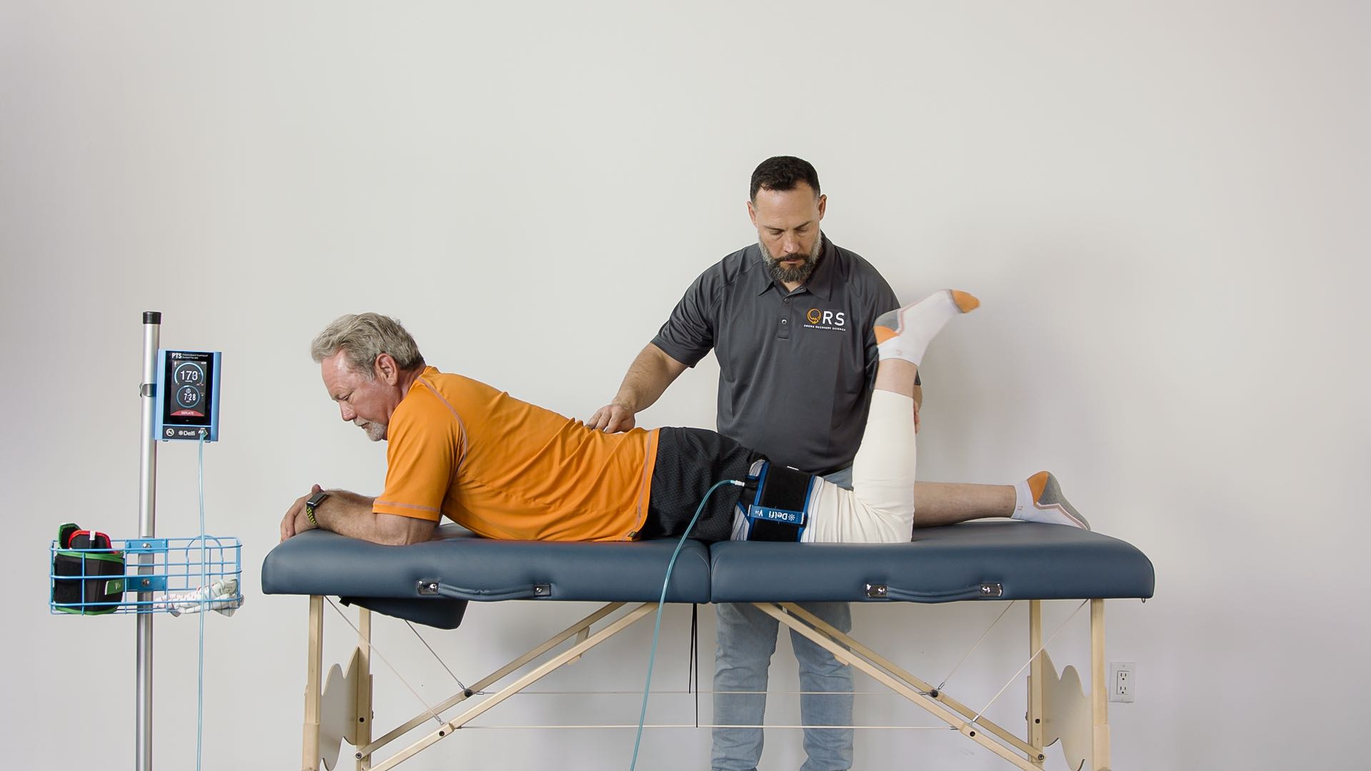
Recently we’ve had a few conversations in our Facebook group, at the NBA Combine, as well as emails regarding how best to manage muscle strains via PBFR. So, we thought we’d put together a brief post centered around this topic. When it comes to this and most other subjects, our chief objective is that we convey the underlying principles driving our decision making and exercise prescription. Having a strong physiologic foundation for our clinical reasoning enables us to apply our knowledge effectively across the broadest range of circumstances. Thus, helping to continue to build this for you will always be our chief focus, because no pre-packaged protocol will ever address comprehensively the individual scenario you will encounter in the clinic. So, let’s dive in further to learn how PBFR can be used for muscle strain treatment.
When one injures a muscle, the body has two primary means of addressing this insult. It involves a delicate interplay between scar and repair. In the context of causing muscular adaptation by lifting heavy weight, our body is quite capable of repairing that damage in a manner that results in growth, or more specifically, increases in strength and hypertrophy. But when one strains a muscle, that insult is often too great to repair fully with muscle tissue, so our scar pathways jump into action to speed up this process. (Järvinen does a beautiful job of describing this process in detail. Järvinen et al. 2005) That can be good as it restores a large amount of function rather quickly, but the resultant reduction in quality tissue affects the muscle’s capacity to perform at a high level. The good news is it looks like from animal research that we can limit that scar via activity. The bad news is that moving the injured tissue hurts, which usually affects how much or how intensely we can move. This was demonstrated elegantly by scientists in the Department of Defense that found forcing a rat to run 1-week after inflicting a large soft tissue injury on their hind limb produced muscle regeneration, the animals who were immobilized primarily produced fibrosis. We as clinicians are unable to force patients with muscle strains to sprint a week after injury and the sedentary state results in the muscle not receiving a strong enough signal to limit scar formation and prioritize muscle regeneration.
The TGF - β superfamily, a scar pathway, is likely the chief driver of the scar response following injury. Myostatin is part of this family, and as presented in the certification course, we can use low load exercise with PBFR to downregulate the myostatin gene very similarly to how heavy lifting will. (Laurentino et al. 2012) Therefore, we would want to knock that gene’s expression down as a means of limiting scar formation and mimicking the "forced running" type of regenerative pathway seen in the rat models. This is one mechanism by which we believe we may help the body to prioritize muscle repair over scarring. In addition to reducing myostatin’s expression, we would also want to maximize our protein synthetic response, as this will optimize our ability to repair rather than scar muscle. Certainly, we know that we can achieve this via PBFR. (Fry et al. 2010; Fujita et al. 2007; Gundermann et al. 2014, for a detailed review read Dr. Bradley Lambert's paper, Lambert et al. 2018) In addition to these two primary mechanisms, there are some other processes we may be able to influence via PBFR to help stimulate the repair of muscle. A key player in the repair process would be the proliferation and differentiation of satellite cells. It looks like the inherent hypoxia created by the damage to the muscle encourages this initially (Jash et al. 2015) and may certainly be further influenced via PBFR exercise.
(Nielsen et al. 2012) Lastly, increases in angiogenesis, (Hunt et al. 2013) and reductions in pain via PBFR may be helpful. (Korakakis et al. 2018; Ellingson et al. 2014; Giles et al. 2017) The latter is something I’ve seen be a very powerful tool in the clinic with this subset of injuries. The pain reductions I’ve seen have been immediate, substantial, and lasting. This provided me the ability to progress to heavier loaded tasks much more rapidly than I anticipated, and thus resulted in shortened courses of care. When it comes to how best to implement PBFR in the management of muscle strains, I would suggest beginning with a load the person can execute without pain or with very tolerable pain. Your goal in this first session is to use PBFR to induce a fatigued state that is surprising to the patient, as they would generally associate this with a bout of heavy lifting that their injury would not allow. Secondly, you will need to assess the person’s tolerance to the cuff pressure, particularly if the injury is underneath the cuff. If it’s too painful then you may need to reduce the pressure, or potentially wait a bit before beginning PBFR. If you’re able to find a load, and your person can tolerate the pressure, then the standard 30/15/15/15 set and rep scheme seems to work well. I’ve found this sufficient to induce fatigue. In some cases, I’ve had to assist the completion of the exercise volume manually, in others I’ve had to increase the volume real time as I may have undershot the load a little.
Either way, if you are able to fatigue the person’s injured muscle, I’ve found they experience an almost immediate reduction in pain. This can obviously be a very valuable thing clinically for a number of reasons. At subsequent visits be sure to re-assess the load you will use prior to the session, and progress it as much as you can with the goal of moving away from PBFR as quickly as possible toward progressively loaded tasks for the injured muscle. You may supplement with PBFR following your heavy session simply to ensure you’ve achieved a large enough protein synthetic response to aid the repair via quality tissue.
Oh yeah and get your protein!! #leftbrisket
Thank you Kyle Kimbrell, MPT for the write up on PBFR for muscle strain treatment.
- Ellingson, L. D., Koltyn, K. F., Kim, J.-S., & Cook, D. B. (2014). Does exercise induce hypoalgesia through conditioned pain modulation? Psychophysiology, 51(3), 267–276.
- Fry, C. S., Glynn, E. L., Drummond, M. J., Timmerman, K. L., Fujita, S., Abe, T., … Rasmussen, B. B. (2010). Blood flow restriction exercise stimulates mTORC1 signaling and muscle protein synthesis in older men. Journal of Applied Physiology, 108(5), 1199–1209.
- Fujita, S., Abe, T., Drummond, M. J., Cadenas, J. G., Dreyer, H. C., Sato, Y., … Rasmussen, B. B. (2007). Blood flow restriction during low-intensity resistance exercise increases S6K1 phosphorylation and muscle protein synthesis. Journal of Applied Physiology, 103(3), 903–910.
- Giles, L., Webster, K. E., McClelland, J., & Cook, J. L. (2017). Quadriceps strengthening with and without blood flow restriction in the treatment of patellofemoral pain: a double-blind randomised trial. British Journal of Sports Medicine, 51(23), 1688–1694.
- Gundermann, D. M., Walker, D. K., Reidy, P. T., Borack, M. S., Dickinson, J. M., Volpi, E., & Rasmussen, B. B. (2014). Activation of mTORC1 signaling and protein synthesis in human muscle following blood flow restriction exercise is inhibited by rapamycin. American Journal of Physiology-Endocrinology and Metabolism, 306(10), E1198–E1204.
- Hunt, J. E. A., Galea, D., Tufft, G., Bunce, D., Ferguson, R. A. (2013). Time course of regional vascular adaptations to low load resistance training with blood flow restriction. Journal of Applied Physiology, 115(3), 403–411.
- Järvinen, T. A. H., Järvinen, T. L. N., Kääriäinen, M., Kalimo, H., & Järvinen, M. (2005). Muscle injuries: biology and treatment. The American Journal of Sports Medicine, 33(5), 745–764. Jash, S., & Adhya, S. (2015). Effects of Transient Hypoxia versus Prolonged Hypoxia on Satellite Cell Proliferation and Differentiation In Vivo. Stem Cells International, 2015, 961307.
- Korakakis, V., Whiteley, R., & Epameinontidis, K. (2018). Blood Flow Restriction induces hypoalgesia in recreationally active adult male anterior knee pain patients allowing therapeutic exercise loading. Physical Therapy in Sport: Official Journal of the Association of Chartered Physiotherapists in Sports Medicine, 32, 235–243.
- Lambert, Bradley S., et al. "Blood Flow Restriction Therapy for Stimulating Skeletal Muscle Growth: Practical Considerations for Maximizing Recovery in Clinical Rehabilitation Settings." Techniques in Orthopaedics 33.2 (2018): 89-97.
- Laurentino, G. C., Ugrinowitsch, C., Roschel, H., Aoki, M. S., Soares, A. G., Neves, M., Jr, … Tricoli, V. (2012). Strength training with blood flow restriction diminishes myostatin gene expression. Medicine and Science in Sports and Exercise, 44(3), 406–412.
- Mason, M. J. S., Owens, J. G., & Brown, L. W. J. (2018). Blood Flow Restriction Training: Current and Future Applications for the Rehabilitation of Musculoskeletal Injuries. Techniques in Orthopaedics, 33(2), 71.
- Nielsen, J. L., Aagaard, P., Bech, R. D., Nygaard, T., Hvid, L. G., Wernbom, M., … Frandsen, U. (2012). Proliferation of myogenic stem cells in human skeletal muscle in response to low-load resistance training with blood flow restriction. The Journal of Physiology, 590(17), 4351–4361.


