If you’ve read our previous blogs on the use of Personalized Blood Flow Restriction (PBFR) for post-surgical conditions, the aging individual or listened to our podcast on “the muscle dump”, you’ve heard us talk about anabolic resistance. We firmly believe you need to define and understand the problem to work toward a solution. Anabolic resistance is one of those “problems” clinicians should understand because it affects every patient that is in a period of disuse (ie...almost all of our patients), happens very quickly after injury or surgery and until now clinicians have had almost no tools to combat it.
What is Anabolic Resistance?
The simple answer is that anabolic resistance is an alternative term (maybe a better one) for disuse atrophy. Elaborating, anabolic resistance is the result of periods of disuse that signal skeletal muscle to waste away. This wasting is thought to be the result of both the amplification of catabolic signaling and reduced anabolic signaling.
This includes but may not be limited to:
- Decreased muscle protein synthesis (MPS)
- Decreased muscle protein synthesis (MPS) refers to a decrease in the rate at which new proteins are created within muscle cells. Decreased MPS may be caused by a variety of factors, including aging muscle, inactivity, and certain medical conditions.
- Increased catabolic markers (signs for muscle protein breakdown): Myostatin, MuRF1, and MAFBx
- Increased catabolic markers, such as myostatin, MuRF1, and MAFBx, have been associated with muscle loss and weakness. Additionally, these markers have been linked to other conditions that lead to muscle wasting, such as cancer and chronic inflammation. While more research is needed to fully understand the role of these markers in muscle loss, it is clear that they are involved in the process.
With the end result of:
- Decreased muscle cross sectional area (CSA)
- Decreased muscle strength
The term anabolic resistance was coined due to the fact that individuals under conditions of limb immobilization displayed a reduced rate of muscle protein synthesis in response to the ingestion of protein. Wall et al examined healthy individuals who were immobilized and forced into non-weight bearing (NWB) on one leg for 2 weeks. This resulted in close to a 30% reduction in the muscle protein synthetic response to protein ingestion. (Wall et al. 2013) The effect of this on the quad was an 8.4% decrease in CSA and a 23% decrease in strength. While it is widely accepted that this reduction in protein synthesis plays a major role in the effects of disuse on muscle, the expression of the catabolic pathways Myostatin, MuRF1, and MAFBx have also been noted in response to disuse in healthy subjects. (Wall et al. 2014) Difficulties with measurement make it more challenging to quantify their impact, and pathology may further amplify it.
Unfortunately, it’s not just complete disuse or immobilization that leads to this blunted muscle protein synthetic response. Reductions in activity can lead to similar changes. Allowing healthy young men to ambulate, but significantly reducing their daily step count by 90% for one week led to a 27% decrease in muscle protein synthesis rate and a 3-fold increase in myostatin. (Shad et al. 2019) Similarly, in healthy elderly individuals, a two week reduction in daily steps resulted in a 26% decrease in protein synthesis and a 4% loss of fat-free leg mass or muscle mass. (Breen et al. 2013)
The prevalence of persistent weakness following injury or surgery is evidence that too often rehab exercise isn't stressful enough to restore a person's skeletal muscle mass and potentially due to inadequate stimulation of muscle protein synthesis. This may be the most apparent in the post ACL reconstruction population. Gumucio et al demonstrated that knee extensor strength after ACL surgery had decreased by 68% in one month and deficits of greater than 20% are still present 6 months after surgery. (Gumucio et al. 2018) This potentially has larger implications for the health of the joint and is potentially a contributor to sarcopenia. (Keays et al. 2010; Lepley 2015)
How Quickly does Anabolic Resistance Happen?
The good news is you’ve got a day! Dirks et al has shown that strength, insulin sensitivity, protein synthesis and catabolic markers are unchanged over a 24 hour period of disuse. (Dirks et al. 2018) The bad news...you might start to lose muscle by day 2. Kilroe et al found that changes in quadriceps and hamstring CSA (1.7 and 1.4% respectively) were evident on MRI at day 2 of immobilization. (Kilroe et al. 2019) The same laboratory found that protein synthesis was reduced by up to 30% with 3 days of immobilization, despite individuals consuming adequate amounts of ingested protein (1.6g/kg/day). (Kilroe et al. 2020) By day 5, there is greater loss of quad muscle size (3.5%) and significant loss of knee extensor strength (9.0%). Additionally, Myostatin, MuRF1, and MAFBx, the catabolic pathways, are significantly elevated. (Wall et al. 2014) In cases of whole body disuse, such as bedrest, as much as 3.1 pounds of muscle can be lost within 1 week. (Dirks et al. 2016) That would equate to six, 8 ounce ribeye steaks worth of muscle!
Now what? How do we combat the problem?
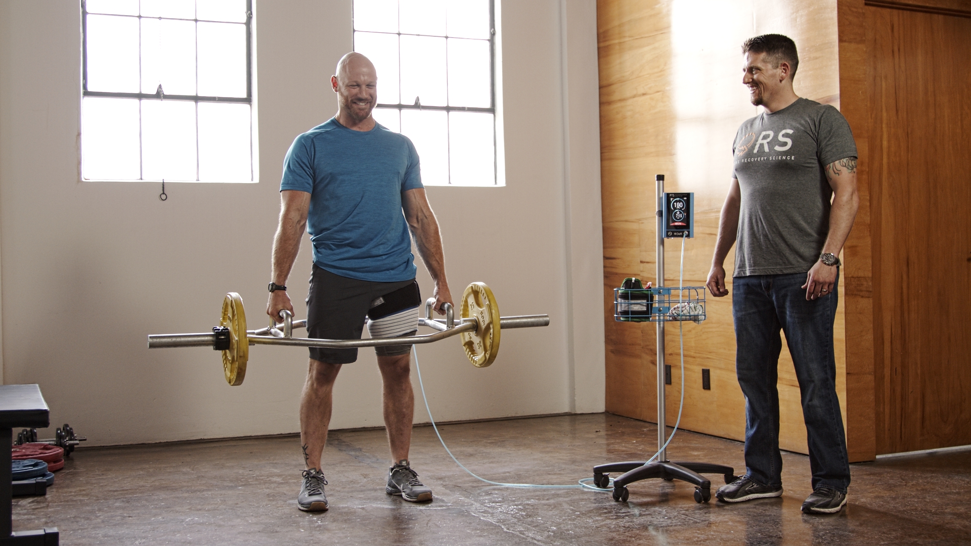
The first step is understanding the problem. As mentioned above, anabolic resistance happens quickly. Significant changes in the response of muscle to nutrient ingestion and markers for catabolism are present within the first 2-5 days. Thus, interventions to reduce these effects and stimulate muscle protein synthesis need to begin as early as possible. The second step involves developing intervention strategies that are effective in slowing catabolic pathways. Applications like passive PBFR, while not ideal, may be our only option at this phase and it may be able to help stave off some of the loss early in the rehab process. A recent paper from Kakehi et al showed that just the application of a tourniquet alone was effective in reducing catabolism. Five rounds of 5 minutes applied twice daily without exercise reduced muscle loss in the thigh and reduced the elevation of MuRF1 during 2 weeks of disuse. The control group who were immobilized and did not receive passive BFR lost significant amounts of muscle and were not able to downregulate MuRF1 (Kakehi et al. 2020) But if the disuse period extends beyond 5-7 days, the need for anabolic stimuli to promote muscle protein accretion becomes more pressing due to the reduced activation of muscle protein synthesis previous studies have identified. In order to do so, we need to add exercise to the tourniquet condition. (Nyakayiru et al. 2019) The addition of neuromuscular electrical stimulation to BFR can be an effective strategy to help bridge the gap back to active exercise. Natsume et al showed that a very low intensity, high frequency application of NMES and BFR increased the size and strength of the quad. (Natsume et al. 2015) Progressing toward multiple, active exercises with BFR in a session is an even better target for making positive muscle changes. Bowman et al showed that 4 exercises per session (SLR, Hip Abduction, Knee Extension, and Knee Flexion), performed twice a week, for 6 weeks significantly increased LE muscle size and strength. (Bowman et al. 2019) Below is a very simple table for how this might progress.
0-5 days | 5-10 days | 10-21 days | |
Passive (5 x 5) | ✓ | ||
NMES + BFR | ✓ | ✓ | |
Active Exercise | ✓ | ✓ |
Goal: Slow catabolism via repeated tourniquet inflations
Goal: Need to get some form of muscle activity to try and increase MPS rates
Goal: Moving toward appropriately loaded exercise w/ high effort to restore lost mass
In the first five days, we’re trying to initiate BFR to slow down the loss of muscle. This may be a time when we can only apply passive tourniquet inflation and 5 rounds of 5 minutes has been shown to be effective. As early as possible, we want to start adding muscle contraction to the equation. A tool like NMES can help bridge the gap if active exercise can’t be achieved or tolerated. For both the passive and NMES applications, a higher frequency of daily or twice a day would be best. As exercises can be added, the frequency can come down to 2 or 3 days per week and multiple exercises per session, such as the 4 in the Bowman study, would be a good target. Ongoing studies with ACL reconstruction, tendon repairs, femur fractures, and total joint replacement should add valuable information on the effectiveness of BFR compared to a work-matched intervention. As usual, we need more research to be completed and published to provide more definitive guidance. For now, make exercise more stressful and don’t forget to eat your brisket or target other forms of high quality protein and leucine.
Additional related podcasts:
- Kilroe SP, Fulford J, Jackman S, et al. Dietary protein intake does not modulate daily myofibrillar protein synthesis rates or loss of muscle mass and function during short-term immobilization in young men: a randomized controlled trial. Am J Clin Nutr. Published online May 29, 2020. doi:10.1093/ajcn/nqaa136
- Kilroe S, Fulford J, Jackman S, van Loon L, Wall B. Temporal Muscle-Specific Disuse Atrophy during One Week of Leg Immobilization. Medicine . 2019;Publish Ahead of Print. doi:10.1249/MSS.0000000000002200
- Shad BJ, Thompson JL, Holwerda AM, et al. One Week of Step Reduction Lowers Myofibrillar Protein Synthesis Rates in Young Men. Med Sci Sports Exerc. 2019;51(10):2125-2134.
- Wall BT, Dirks ML, Snijders T, Senden JMG, Dolmans J, van Loon LJC. Substantial skeletal muscle loss occurs during only 5 days of disuse. Acta Physiol . 2014;210(3):600-611.
- Dirks ML, Wall BT, van de Valk B, et al. One Week of Bed Rest Leads to Substantial Muscle Atrophy and Induces Whole-Body Insulin Resistance in the Absence of Skeletal Muscle Lipid Accumulation. Diabetes. 2016;65(10):2862-2875.
- Dirks ML, Stephens FB, Jackman SR, et al. A single day of bed rest, irrespective of energy balance, does not affect skeletal muscle gene expression or insulin sensitivity. Exp Physiol. Published online April 15, 2018. doi:10.1113/EP086961
- Breen L, Stokes KA, Churchward-Venne TA, et al. Two weeks of reduced activity decreases leg lean mass and induces “anabolic resistance” of myofibrillar protein synthesis in healthy elderly. J Clin Endocrinol Metab. 2013;98(6):2604-2612.
- Wall BT, Snijders T, Senden JMG, et al. Disuse impairs the muscle protein synthetic response to protein ingestion in healthy men. J Clin Endocrinol Metab. 2013;98(12):4872-4881.
- Hughes L, Rosenblatt B, Haddad F, et al. Comparing the Effectiveness of Blood Flow Restriction and Traditional Heavy Load Resistance Training in the Post-Surgery Rehabilitation of Anterior Cruciate Ligament Reconstruction Patients: A UK National Health Service Randomised Controlled Trial. Sports Med. Published online July 12, 2019. doi:10.1007/s40279-019-01137-2
- Natsume T, Ozaki H, Saito AI, Abe T, Naito H. Effects of Electrostimulation with Blood Flow Restriction on Muscle Size and Strength. Med Sci Sports Exerc. 2015;47(12):2621-2627.
- Bowman EN, Elshaar R, Milligan H, et al. Proximal, Distal, and Contralateral Effects of Blood Flow Restriction Training on the Lower Extremities: A Randomized Controlled Trial. Sports Health. Published online January 14, 2019:1941738118821929.
- Keays SL, Newcombe PA, Bullock-Saxton JE, Bullock MI, Keays AC. Factors involved in the development of osteoarthritis after anterior cruciate ligament surgery. Am J Sports Med. 2010;38(3):455-463.
- Lepley LK. Deficits in Quadriceps Strength and Patient-Oriented Outcomes at Return to Activity After ACL Reconstruction: A Review of the Current Literature. Sports Health. 2015;7(3):231-238.